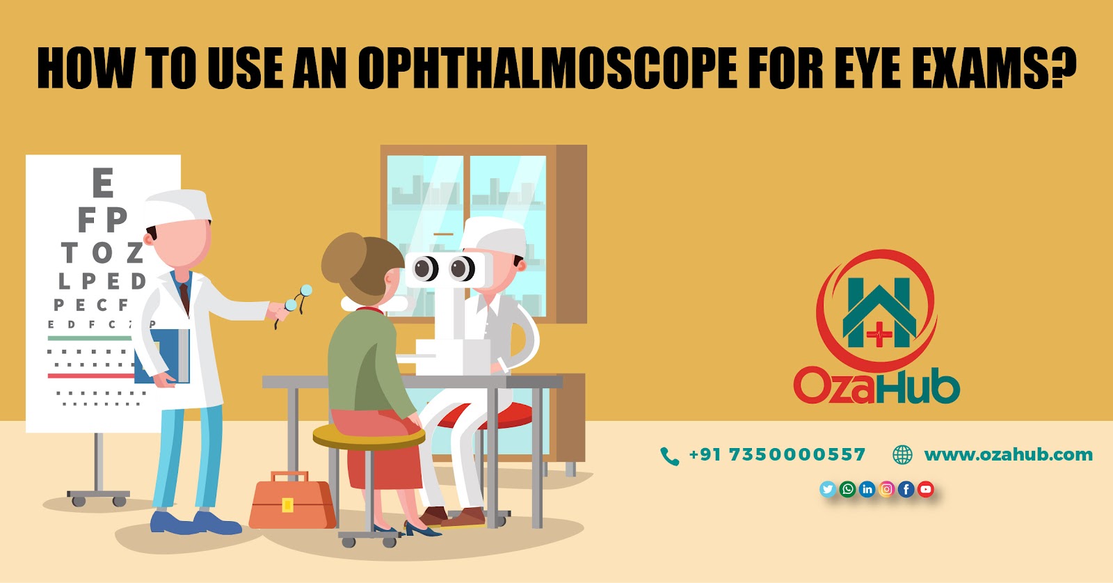
HOW TO USE AN OPHTHALMOSCOPE FOR EYE EXAMSPosted by ozahub on December 18th, 2020 HOW TO USE AN OPHTHALMOSCOPE FOR EYE EXAMS Ophthalmoscopy, also called fundoscopy, refers to the kind of analysis precise to eye inspections that allows a physician to see inner the fundus of the eye. The ophthalmoscope occasionally denoted as a fundoscopy, is one of a crowd of tools used by doctors to inspect the eye. Many of the ophthalmoscopes operated today are what we would denote as pocket ophthalmoscopes. An ophthalmoscope is recognized as a coaxial visual scheme in which the bloc of light concurs with the alliance of sight into the retina. In other words, the ophthalmoscope removes obscurities in a way that allows for a strong fundus inspection notwithstanding the obviously minor dimension of the pupil. It's a normal instrument in any well-supplied therapeutic stock storeroom. There are two kinds of ophthalmoscopes that are being supplied by the Ophthalmoscope suppliers and they are the old-style straight ophthalmoscope and Panoptic or secondary ophthalmoscope. A direct ophthalmoscope produces a standing carbon copy of the midpoint of the retina at about 15x intensification. A PanOptic ophthalmoscope creates an upturned (reversed) copy of the retina in its totality at about 2x to 5x intensification.
The ophthalmoscope being manufactured by any Ophthalmoscope manufacturer should have an opening variety round or sieve knob that allows the ophthalmoscope to be operated for diverse tenacities by adapting how much brightness is permitted through the lens. The Ophthalmoscope should possess a governor control that controls the brightness cause and permits the doctor to physically regulate the illumination of the halogen beam. The Ophthalmoscope should also possess manifold watching lenses that vary by the device being supplied by the Ophthalmoscope suppliers in India. The Ophthalmoscope should possess a focusing wheel that might seem threatening to the new operator. The last feature that an ophthalmoscope should have is a brow rest that lets the eye medic or inspector to wear spectacles while it. How to Use an Ophthalmoscope Before the specialist begins his examination, it is essential to adapt the setting to make it appropriate for testing. The part openly adjacent to the patient should be empty in mandate to allow for movement of the assessor and placing of the scale. The inspection area should have loads of illumination choices as many of the examinations need a dimly lit room. 1. The physician should start by asking the patient to remove any spectacles or contact lenses. The inspector may wear their own remedial eyewear in place. 2. Confirm the ophthalmoscope is in operational order and turn it on. Retain in the notice that a non-working ophthalmoscope may just be out of batteries or need to be charged. 3. Blacken or faint the room. Doctors often use mydriatic droplets to added pupil enlargement. While expanding the eye exploits the watching competencies, it is not essential to use eye droplets in order to carry out the examination. Not watered-down pupils will simply outcome in a lesser field of view. 4. The Doctor should ask the patient to sit motionlessly and center their stare on an exact thing in the examination room. 5. The physician should set the ophthalmoscope light to extreme illumination and turn the knob until one sees a round circle. The Doctor will want this bright light to be white, so he should put the ophthalmoscope lens on the red-free filter.
6. The Physician should start by playing a red reflex test. The determination of the red reflex test is to check for the likeness of dainty from the retina. The nonappearance of this image may indicate the company of an eye complaint. 7. With the ophthalmoscope about six inches from the patient, the doctor should use his right hand and right eye to look through the ophthalmoscope at the patient's right eye. The doctor should adjust the wheel toward the bad or red end of the component until his view is clear and in focus. He should move the ophthalmoscope 15 degrees from the midpoint and look for the red reflex, following it until the retina comes into view. 8. Now that the doctor has found the retina, his next move will vary slightly based on whether he is carrying out direct ophthalmoscopy or indirect ophthalmoscopy. If you are using a direct ophthalmoscope, hinge the ophthalmoscope by slanting the device up, down, left, and right. If the doctor is using the Panoptic ophthalmoscope, he can similarly pivot using very small actions or can ask the patient to look up, down, left, and right. 9. Lasting with the red reflex as the conductor, find the optic disc and turn the lens dial right or left until it comes into focus. The doctor will need to move nearer to the patient because the optic disc generally comes into view when you are one to two inches from the patient's eye. The doctor needs to be close enough to the patient to get a precise opinion. 10. As the doctor examines the fundus of the eye and the optic disc, he should follow the retinal arteries and examine the vascular arcades. In other words, he should follow each vessel as far out to the periphery as he can. 11. The last part of the inspection is an assessment of the macula of the eye. To examine the macula, the doctor must move the ophthalmoscope back several inches and ask the patient to look directly at its light. For all your ophthalmoscope needs please visit Ozahub today. Like it? Share it! More by this author |